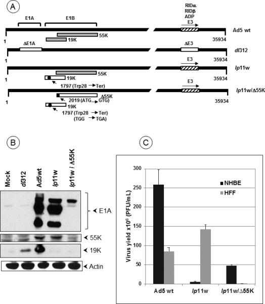
Figure 1. HAdV5 mutants and replication in normal cells.

(A). Diagrammatic illustration of HAdV5 mutants. The top panel shows the proteins expressed from early regions E1B and E3. Mutants lp11w and lp11w/Δ55K express wt E1A and E3 regions while the control mutant dl312 contain deletions in these regions. The mutations within the E1B region are indicated. (B). Expression of E1A and E1B proteins by HAdV5 mutants. (C). Replication of HAdV5 mutants in normal human epithelial (NHBE) and fibroblast (HFF) cells. The virus yield from infected NHBE and HFF cells were determined by plaque assay on 293 cells.
