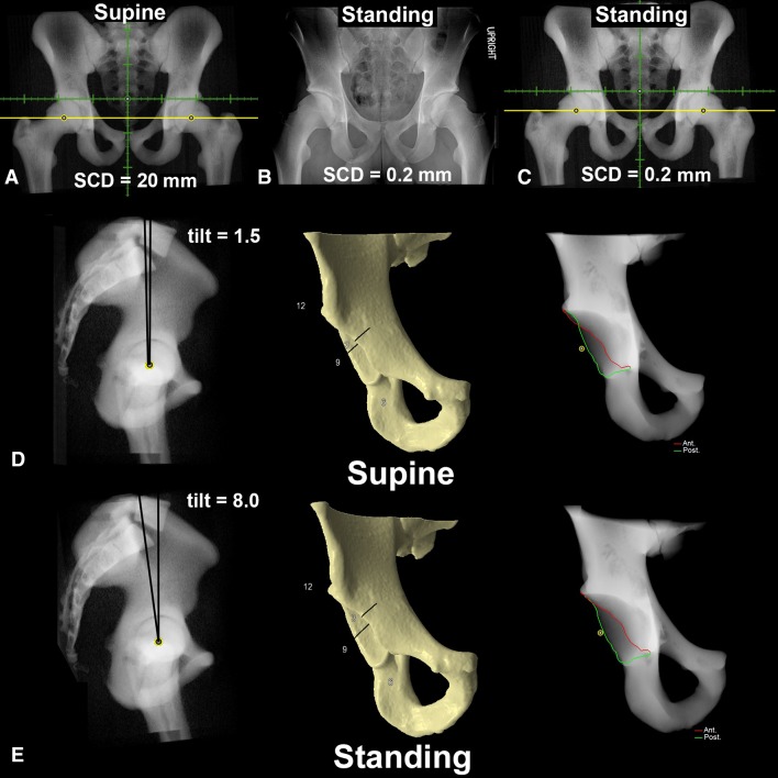
Fig. 1A–E.
Patient example demonstrating the supine virtual radiograph (A) reconstructed from the CT scan. (B) The standing AP pelvic radiograph demonstrated a SCD of 0.2 mm. The standing virtual radiograph (C) was created by increasing posterior pelvic tilt (D–E) by 6.5°. (D) Supine pelvic radiograph demonstrated a positive crossover sign, which is eliminated with standing radiographic evaluation (E).

