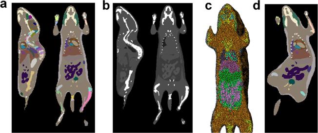
Fig. 6.
Voxelization and tetrahedralization of the atlas. a The atlas is voxelized into a labeled image with a 0.2-mm resolution. The labeled image is shown in sagittal and coronal slices with pseudo color. b The labeled image is converted into pseudo CT by assigning different organs with appropriate Hounsfield values. c The labeled image is converted into tetrahedral mesh. d A voxelized label image of an arbitrary body pose and weight.

