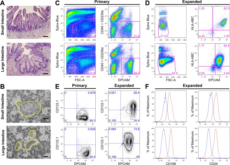
Fig 1. In vitro expanded primary epithelial cells from human fetal small and large intestine express stem cell markers.
A-F, top panels: small intestine, bottom panels: large intestine (A) Hematoxylin and eosin paraffin sections of normal fetal intestine at 23 weeks of gestation. (B) Intestinal cells expanded on the feeder layer (yellow outline). (C-F) Representative analyses of primary and expanded cells at passages 4, 5 or 6 with the percentage of gated live cells provided. (C) Flow cytometric analysis (dot plots) of freshly isolated primary intestinal cells. Gating strategy identifies total live cells (left panels) and EPCAM+ cells, excluding blood cells (CD45+CD235a+) (right panels). (D) Flow cytometric analysis of expanded cells. Gating strategy identifies total live cells (left panels) and EPCAM+ cells, excluding rodent feeder layer cells (HLA−) (right panels). (E) Flow cytometric analysis (5% probability contour plots) of primary and expanded intestinal cells, stained with EPCAM and CD133.1. (F) Expanded intestinal cells also express other putative stem cell markers. Histograms represent unstained control (blue) and stained with CD166 or CD24 (red) of live EPCAM+ cells. Scale bars = 100μm for panel A and B.

