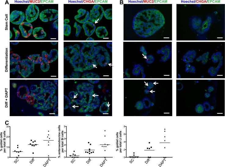
Fig 5. Immunofluorescent staining of intestinal organoids confirms multipotentiality and differentiation potential.
(A) SI and (B) LI organoids from one paired sample stained for epithelial marker, EPCAM (green), goblet cell marker, MUC2 (red) and enteroendocrine marker, CHGA (red) treated with SC media, Differentiation media (Diff) or Diff media + DAPT. Counterstain, Hoechst 33342. White arrows indicate positive cells. Scale bar = 50μm. (C) Quantification of percent goblet or enteroendocrine cells of total cells counted per field of view. Not enough enteroendocrine cells were present in the organoids derived from LI for quantification. This paired set serves as a representative of two sets examined.

