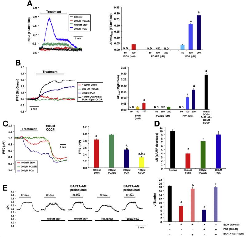
Figure 4.
High concentrations of ethanol and POA induce sustained elevation of [Ca2+]i, impaired mitochondrial function, and decreased cAMP levels in Capan-1 PDECs. (A) Representative traces and summary data of the ΔRatiomax show the effect of ethanol, POAEE, and POA on [Ca2+]i. Ethanol (100 mmol/L) induced a small, sustained elevation of [Ca2+]i, whereas 100 to 200 μmol/L POA induced a significantly higher increase in [Ca2+]i. aP < .05 vs 100 mmol/L ethanol. (B) Ethanol and POA induced significant and irreversible depletion of (ATP)i. Deoxyglucose/iodoacetic acid (DOG/IAA; glycolysis inhibition) and CCCP (inhibition of mitochondrial ATP production) served as control. (C) Representative traces and summary data of changes in the mitochondrial membrane potential [(ΔΨ)m]. Ethanol (100 mmol/L) induced a moderate decrease in (δΨ)m, whereas 200 μmol/L POA had a more prominent effect. CCCP induced a further decrease in (δΨ)m after treatment with POA. (D) Summary data for cAMP measurements. A total of 100 mmol/L ethanol and 200 μmol/L POAEE significantly decreased forskolin-stimulated cAMP production. (E) Ca2+ chelation abolished the inhibitory effect of ethanol and POA on intracellular pH recovery after luminal CI− readdition. For all conditions, n = 3–5/group. aP < .05 vs control; bP < .05 vs 100 mmol/L ethanol; cP < .05 vs 200 μmol/L POA. N.D., not detected.

