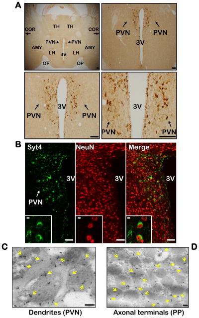
Figure 1. Syt4 distribution in hypothalamic PVN.
A. Distribution of Syt4 in the hypothalamic PVN. Immunohistochemical staining of Syt4 in hypothalamic sections across the PVN was examined under a light microscope. Scale bar=100 μm.
B. Neuronal expression of Syt4 in the PVN. PVN sections were co-immunostained for Syt4 (green) and neuronal marker NeuN (red). Co-localization of two fluorescent signals within the same cells indicates Syt4 expression in neurons. Scale bar=50 μm. Inserts: Intracellular distribution of Syt4 in neurons by co-immunostaining at high magnification (insert scale bar=5 μm).
C&D. Syt4 immunogold labeling in hypothalamic PVN (C) and posterior pituitary (D) sections were examined by electron microscopy. Yellow arrows indicate Syt4 immunogold labeling. Scale bar=100 nm.
A–D: All experimental mice were adult males, chow-fed, and in C57BL/6 background. AMY: amygdala, COR: cortex, LH: lateral hypothalamus, OP: optic tract, PVN: paraventricular nucleus, PP: posterior pituitary, TH: thalamus, 3V: third ventricle.

