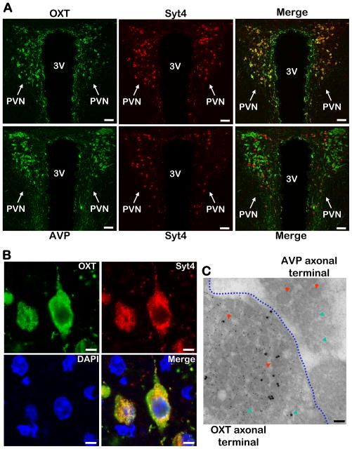
Figure 4. Co-localization of Syt4 with OXT in hypothalamic PVN.
A. OXT (upper panels, green) or AVP (lower panels, green) in the PVN was co-immunostained with Syt4 (all panels, red) and merged to display their co-localization (indicated by yellow color). Scale bar=50 μm.
B. High-magnification images of Syt4 (red) and OXT (green) co-immunostaining. Yellow color in merged images indicates intracellular co-localization of Syt4 and OXT. DAPI staining (blue) revealed nuclei of all cells in the sections. Scale bar=5 μm.
C. OXT and Syt4 co-immunogold labeling in OXT vs. AVP axonal terminals. The posterior pituitary from normal C57BL/6 mice were sectioned and co-immunogold labeled with OXT (small particles) and Syt4 (large particles). The image represents a junction region that contains both OXT axonal terminals and AVP axonal terminals (separated by a blue dotted line). Red arrows indicate dense-core vesicles, and green arrows indicate micro-vesicles. Scale bar=100 nm.
A–C: All experimental mice were adult males, chow-fed, and in C57BL/6 background.

