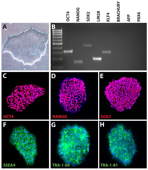
Figure 2. Characterization of undifferentiated hPSCs.
hPSCs displayed a typical undifferentiated morphology, including tightly packed colonies of cells and clearly defined edges (A). RT-PCR analysis demonstrated the expression of characteristic pluripotency makers in hPSCs, while lacking mesodermal, endodermal and ectodermal markers (B). Immunocytochemistry further demonstrated widespread expression of pluripotency-associated transcription factors (C–E) as well as cell surface markers (F–H). See Tables 1 and 2 for a listing of primers and antibodies used for RT-PCR and immunocytochemistry.

