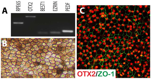
Figure 4. Differentiation of hPSCs to Retinal Pigmented Epithelium.

hPSC-derived RPE-like cells expressed typical RPE associated markers when screened by RT-PCR (A). Under brightfield microscopy, these cells displayed proper morphological features distinct to RPE, including a hexagonal shape and areas of pigmentation (B). Immunocytochemical analysis revealed the features typical of the retinal pigmented epithelium (C).
