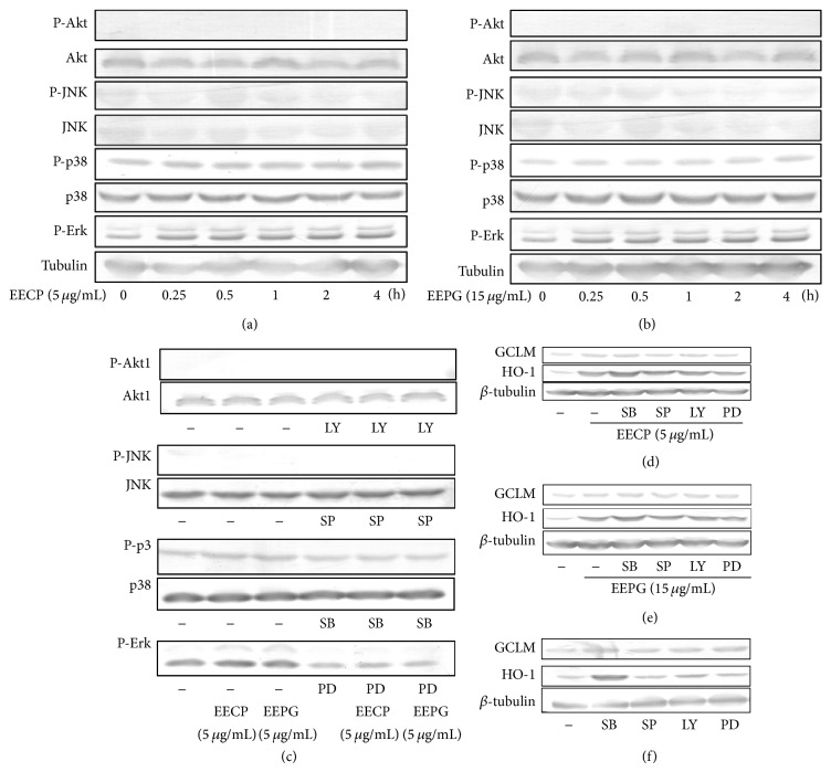
Figure 6.
EECP and EEPG mediate antioxidant genes expression mainly through p38/P-p38 and Erk/P-Erk pathways. ((a), (b)) RAW cells were treated with indicated concentrations of EECP and EEPG for following lengths of time, respectively. Then, the cells were harvested with NP40 and cytoplasmic proteins were extracted. Expressions of Akt, phosphorylated Akt, JNK, phosphorylated JNK, p38, phosphorylated p38, and phosphorylated Erk were determined by western blot. (c) RAW cells were pretreated with or without inhibitors (LY294002, 20 μM; SP600125, 20 μM; SB203580, 30 μM; PD98059, 20 μM) for 0.5 h. After that, cells were cultured in the presence or absence of EECP and EEPG on the indicated concentrations for 1 h and the cytoplasmic protein were collected by NP40. Examining the expressions of Akt, phosphorylated Akt, JNK, phosphorylated JNK, p38, phosphorylated p38, and phosphorylated Erk by western blot. ((d)–(f)) RAW264.7 cells were pretreated with or without inhibitors (LY294002, 20 μM; SP600125, 20 μM; SB203580, 30 μM; PD98059, 20 μM) for 0.5 h, followed by culturing with or without EECP or EEPG for 5 h. Then, the medium were removed and cultured RAW264.7 cells with fresh medium for further 4 h. At the harvest time, protein was collected and western blot was used to detect the expression of TrxR1, HO-1, GCLM and β-tubulin. β-tubulin was used as a protein loading control for each lane.

