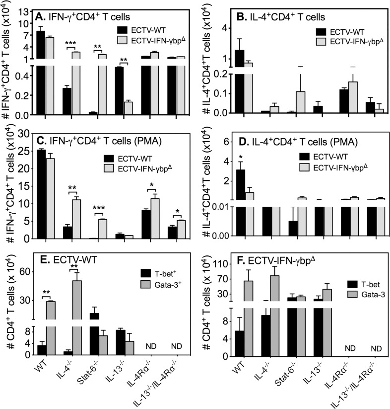
Fig 5. Numbers of CD4 T cells expressing IFN-γ, IL-4, T-bet or GATA-3.
Mice (n = 5–10/group) were infected with ECTV-WT or ECTV-IFN-γbpΔ, sacrificed on day 7 p.i. and splenocytes used for intracellular staining for IFN-γ, IL-4, T-bet or GATA-3 without stimulation (A and B) or with PMA + ionomycin stimulation (C and D). Shown are means of absolute numbers of IFN-γ+CD4 T cells with no stimulation (A) or PMA + ionomycin stimulation (C), IL-4+CD4 T cells with no stimulation (B) or PMA + ionomycin stimulation (D). Also shown are means of absolute numbers of T-bet+ and GATA-3+ CD4 T cells following ECTV-WT (E) or ECTV-IFN-γbpΔ (F) infection. Two-way ANOVA followed by Fisher’s LSD test for significance was used. For panel A, numbers of IFN-γ+CD4 T cells in WT mice for both viruses were significantly higher (p<0.0001) compared to numbers in all GKO strains. In IL-4−/− and STAT-6−/− mice, IFN-γ+CD4 T cell numbers generated by ECTV-IFN-γbpΔ infection were significantly higher (p = 0.0005 and p = 0.0013, respectively) compared to WT virus infection. In IL-13−/− mice, IFN-γ+CD4 T cell numbers generated by WT virus infection were significantly higher (p = 0.0034) compared to ECTV-IFN-γbpΔ infection. For panel B, numbers of IL-4+CD4 T cells in WT mice for both viruses were significantly higher (p<0.01) compared to numbers in all GKO strains. No other significant differences were found. For panel C, numbers of IFN-γ+CD4 T cells in WT mice for both viruses were significantly higher (p<0.0001 in ECTV-WT and p < 0.001 in ECTV-IFN-γbpΔ) compared to numbers in all GKO strains. In IL-4−/−, STAT-6−/− IL-4Rα−/− and IL-13−/− / IL-4Rα−/− mice, IFN-γ+CD4 T cell numbers generated by ECTV-IFN-γbpΔ infection were significantly higher compared to WT virus infection. For panel D, numbers of IL-4+CD4 T cells in WT mice for ECTV-WT virus was significantly higher (p<0.05) compared to numbers in all GKO strains. In WT mice, IL-4+CD4 T cell numbers generated by WT virus infection were significantly higher (p<0.05) compared to ECTV-IFN-γbpΔ infection. For panel E, T-bet+ CD4 T cell numbers were significantly increased (p<0.01) in STAT-6−/− and IL-13−/− mice compared to WT animals. For panel F, no significant differences were found between strains. Data shown for panels A and B are from one of two independent experiments with similar results. Data shown for panels C-F are from one experiment. *, p < 0.05; **, p < 0.01; ***, p < 0.0001.

