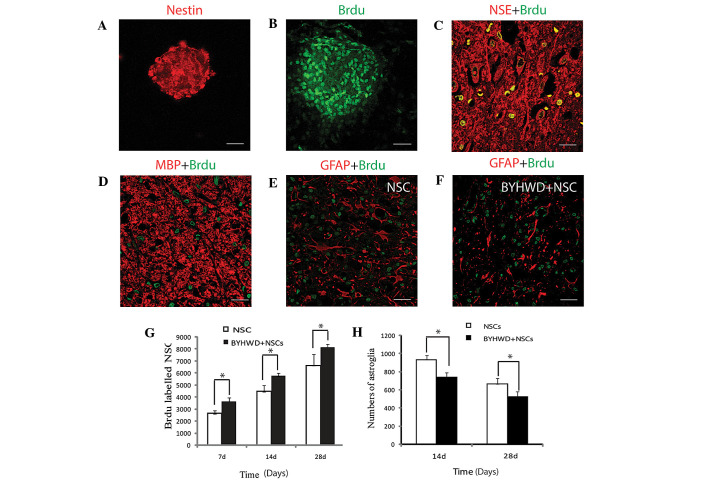
Figure 1.
Characterization and differentiation of the cultured isolated NSCs from the rat embryo neural tube. (A) Cultured NSCs grew into neurospheres in the culture dish, expressing the neural stem cell marker, nestin. (B) NSCs continued to proliferate in the in vitro culture, as shown by the incorporation of BrdU (green). (C) Transplanted NSCs in the injured spinal cord differentiated into neurons, labeled with the neuron marker, NSE (red) and BrdU (green). (D) Transplanted NSCs differentiated into oligodendrocytes, expressing MBP and BrdU, and incorporated into neural networks. (E and F) BYHWD treatment exerted a suppressive effect on astrogliosis in the spinal cord injury (SCI) site, as compared with NSC treatment alone. (G) BYHWD treatment was shown to promote NSC survival at the SCI site at the three examined time points (*P<0.05; n=36). (H) BYHWD treatment was shown to inhibit astrogliogenesis in the surrounding SCI site at the two examined time points (*P<0.05; n=36). BYHWD, Buyang Huanwu decoction; NSC, neural stem cells; BrdU, 5-bromo-2-deoxyuridine; GFAP, glial fibrillary acidic protein; MBP, myelin basic protein; NSE, neuron specific enolase. Scale bars, 50 μm (A and C), 30 μm (B) and 100 μm (D, E and F).

