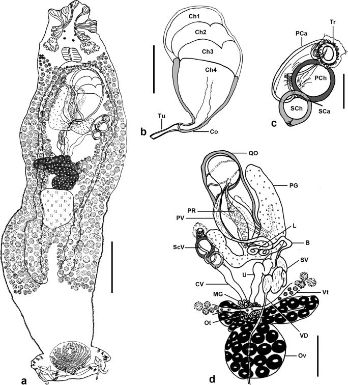
Figure 1.
Pseudorhabdosynochus jeanloui n. sp. from Paranthias colonus. a, composite, mainly from holotype, Hoyer, tegumental scales only on edges, ventral view. b, quadriloculate organ, holotype, Hoyer, ventral view. Ch1-4, chambers; Co, cone; Tu, tube. c, sclerotized vagina, ventral view. Tr, trumpet; PCa, primary canal; PCh, primary chamber; SCa, secondary canal; SCh, secondary chamber. d, male and female organs, testis not represented, paratype, carmine, dorsal view. VD, vas deferens; SV, seminal vesicle; B, bend; L, loops; PV, deferent enlarged posterior vesicle; PR, prostatic reservoir; PG, prostatic gland; QO, quadriloculate organ; Ov, ovary; Ot, ootype; Vt, vitelline ducts; MG, Mehlis’s glands; U, uterus; CV, unsclerotized canal from secondary chamber of vagina to ootype; ScV, sclerotized vagina. Scale bars: a, 100 μm; b, d, 40 μm; c, 20 μm.

