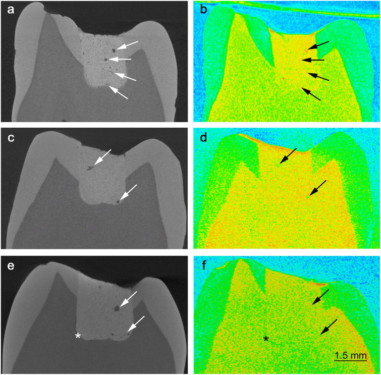
Figure 1. Extracted teeth restored with GIC.
Pores and cracks are better visible in the X-ray images due to better resolution (a, c, e). Poor adaptation (*) of the more viscous restorative cement (Poly) at the bottom of the cavity is observed (e), while this problem is less evident for the less viscous cement (a, c). The presence of liquid inside or adhered to the internal walls of some of the larger pores is evident (in red, due to the higher attenuation coefficient of hydrogen) in the neutron image (b). The neutron images also suggest that interconnecting pores or cracks are filled with liquid (b, f), while some of the larger pores seem to be empty (d).

