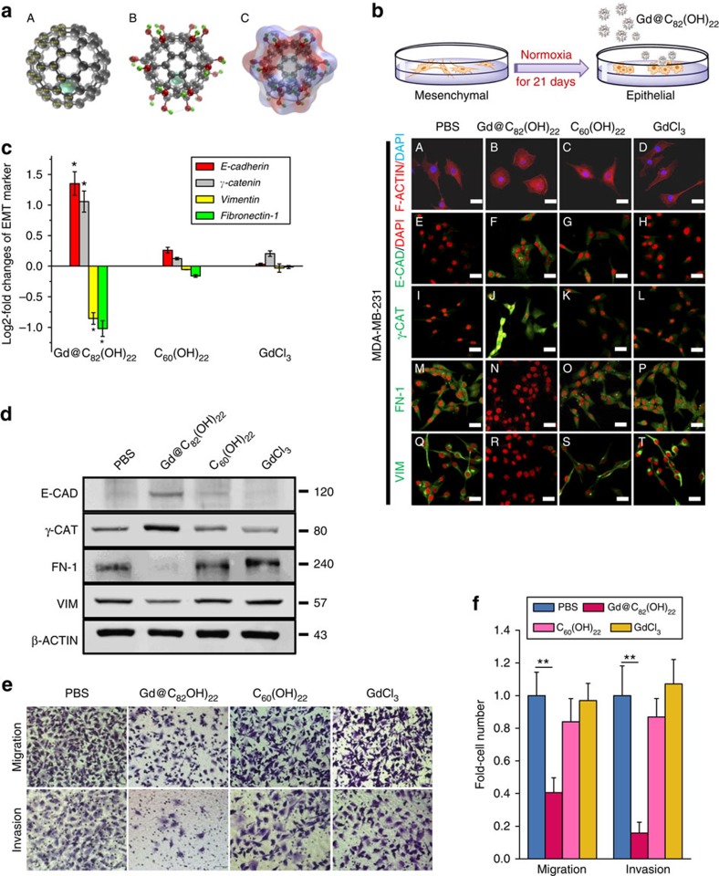
Figure 1. Treatment with Gd@C82(OH)22 impeded EMT, invasion and metastasis of TNBC cells in vitro.
(a) Schematic drawing of Gd@C82(OH)22 nanoparticles. (A) Atomic structure of Gd@C82, which has a C2v point group symmetry. Mülliken atomic charges of symmetrically unique atoms are marked. (B) Atomic structure of Gd@C82(OH)22. The dashed line indicates an intramolecular hydrogen bonds between hydrogen and carbon atoms. (C) Atomic structure of Gd@C82(OH)22 with colour on the molecular surface showing approximated electrostatic potential. (b) MDA-MB-231 cells were treated with PBS (A, E, I, M, Q), Gd@C82(OH)22 (B, F, J, N, R), C60(OH)22 (C, G, K, O, S) or GdCl3 (D, H, L, P, T) (all 50 μM) for 21 days. Phalloidin staining of F-ACTIN cytoskeleton (A–D) of cells in monolayer culture (scale bar, 12.5 μm) was visualized. (c) mRNA levels of EMT markers (E-cadherin, γ-catenin, Vimentin and Fibronectin-1) were analysed by qPCR (mean±s.e.m., n=3 each). *P<0.05 (ANOVA, Tukey’s post-hoc test). Protein levels of E-CADHERIN, γ-CATENIN, VIMENTIN and FIBRONECTIN-1 were detected by immunofluorescence staining (b) and western blot (d). Scale bar, 25 μm. (e,f) Cell migration and invasion were examined using trans-well cell culture chambers and Matrigel-coated ones (mean±s.e.m., n=6 each). **P<0.01 (one-way ANOVA, Tukey’s post-hoc test).

