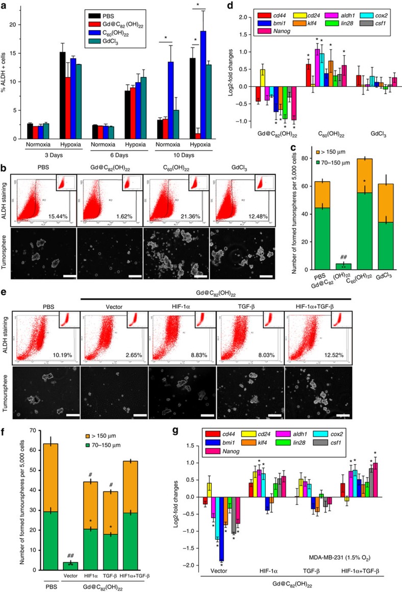
Figure 9. Gd@C82(OH)22 efficiently eliminated breast CSC population through TGF-β and HIF-1a.
MDA-MB-231 cells were maintained in PBS, Gd@C82(OH)22, C60(OH)22 or GdCl3 under hypoxia for 10 days for ALDEFLUOR (a) and tumoursphere formation assay (b) (scale bar, 100 μm) (mean±s.e.m., n=3 each). *P<0.05 (two-way ANOVA, Bonferroni’s post-hoc test). (c) The tumourspheres count (mean±s.e.m., n=3 each). To tumourspheres of 70–150 μm, *P<0.05 and **P<0.01; to tumourspheres of > 150 μm, #P<0.05 and ##P<0.01 (one-way ANOVA, Tukey’s post-hoc test). (d) The mRNA levels of CSC markers were analysed by qPCR (mean±s.e.m., n=3 each). **P<0.01 (one-way ANOVA, Tukey’s post-hoc test). (e) Tumoursphere formation assay after MDA-MB-231 cells were transfected with HIF-1α expressing plasmid and/or supplemented with 20 ng ml−1 TGF-β and cultured under hypoxia for 10 days (scale bar, 12.5 μm). (f) The tumourspheres count (mean±s.e.m., n=3 each). To tumourspheres of 70–150 μm, *P<0.05 and **P<0.01; to tumourspheres of >150 μm, #P<0.05 and ##P<0.01 (two-way ANOVA, Bonferroni’s post-hoc test). (g) The mRNA levels of CSC markers were analysed by qPCR (mean±s.e.m., n=3 each). *P<0.05 (two-way ANOVA, Bonferroni’s post-hoc test).

