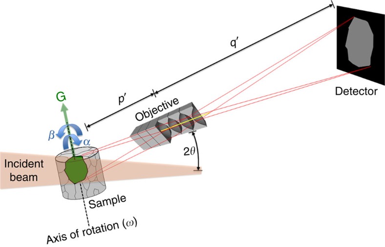
Figure 1. Principle of dark-field X-ray microscopy.
A monochromatic beam from a synchrotron source illuminates the sample. An embedded crystalline element (for example, a grain or domain) of choice (green) is aligned such that the beam is diffracted. The objective magnifies the diffracted beam by a factor M=q′/p′ and generates an inverted 2D projection of the grain. Through repeated exposures during a 360° rotation of the element around an axis parallel to the diffraction vector, G, several 2D projections of the grain are obtained from various angles30. A 3D map is then obtained by combining these projections using reconstruction algorithms similar to those developed for CT scanning32. If the lattice of the crystalline element exhibits an internal orientation spread, this procedure is repeated for a number of sample tilts, indicated by the angles α and β. Using a compound refractive lens18,29 as the objective enables one to enlarge or reduce the spatial resolution and field of view within the sample by varying the number of individual lenses and adjusting p′ and q′ correspondingly. The diffraction angle 2θ is typically 10–30°.

