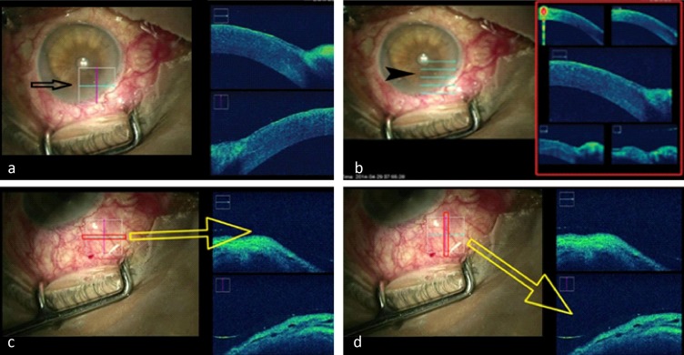
Figure 2. .
Types of scans available: left side (black arrow) shows a cube scan (a) and the right side (arrow head) shows 5-line raster scans (b); horizontal (c) and vertical (d) scans of the regions of interest and the corresponding live OCT imaging. Cross-section at the level of the blue horizontal line is shown at the top right OCT image (c) and the red vertical line is shown at the bottom right OCT image (d).

