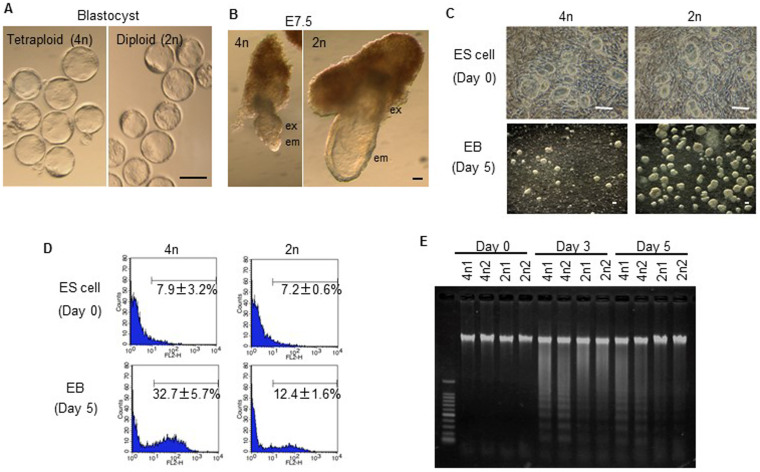
Figure 1. Tetraploid ES cells show apoptosis after differentiation induction.
(A) Tetraploid (4n) and diploid (2n) embryos are morphologically similar at the blastocyst stage. (B) Epiblast-derived embryonic tissue is retarded in tetraploid embryos at E7.5. (C) Undifferentiated tetraploid and diploid ES cells appear morphologically similar (top). Following differentiation induction, tetraploid ES cells form poorly developed EBs compared to diploid ES cells (bottom). (D) FACS analysis for active caspases before and after differentiation induction. The numbers show the percentages of caspase-positive cells. (E) Detection of apoptosis using the DNA ladder method for tetraploid ES cells lines, 4n1 and 4n2 and diploid ES cell lines, 2n1 and 2n2.

