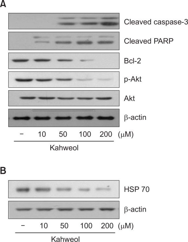
Fig. 3.
Effect of kahweol on the expression of apoptosis-related proteins (A) and HSP70 (B) in HT-29 cells. Cells were treated with the indicated concentrations of kahweol for 16 h, and then the expression levels of cleaved caspase-3, poly (ADP-ribose) polymerase (PARP), Bcl-2, phosphorylated Akt (p-Akt), total Akt, and HSP70 were determined by Western blot analysis. β-actin was used as a loading control.

