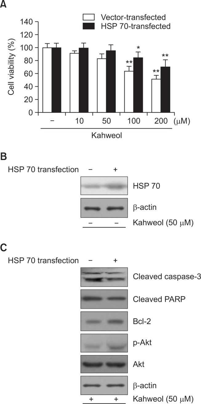
Fig. 5.
Effect of HSP70 overexpression on kahweol-induced apoptitic cytotoxicity in HT-29 cells. A. Cells were transiently transfected with HSP70, and then incubated with the indicated concentrations of kahweol for 24 h. Cell viability was measured by MTT. B. The expression level of HSP70 was determined by Western blot analysis. Compared with cells transfected with vehicle (control), HSP70 expression was significantly elevated in HSP70-transfected cells. C. HSP70 overexpression significantly reduced the expression of caspase-3 and PARP and increased the expression of Bcl-2 and p-Akt. β-actin was used as a loading control. Results are expressed as the mean ± SD of three or more independent experiments. *p<0.05, and **p<0.01 vs. control.

