Figure 1.
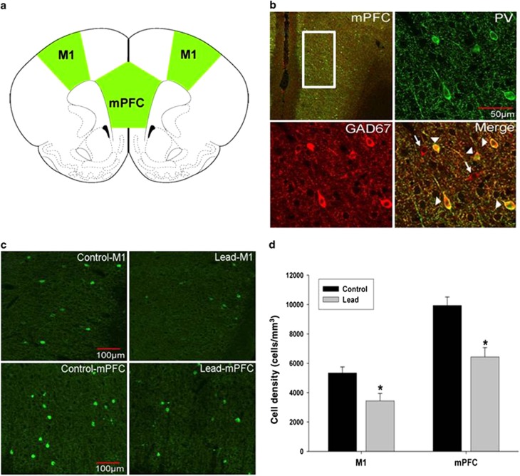
Parvalbumin-positive cell density in the primary motor cortex (M1) and medial prefrontal cortex (mPFC) of control and Pb2+ treated rats. (a) Rat atlas depictions of frontal cortex regions traced in green and used for PVGI cell counting. (b) Parvalbumin-positive interneurons (PV, green) in the mPFC co-labeled with GAD67 (red). However, not all GAD67-labeled cells co-labeled with PV consistent with the fact that only a portion of GABAergic interneurons are PV positive. Arrowheads point to PV and GAD67 co-labeled cells. Arrows point to GAD67-positive cells that do not co-label with PV. (c) Representative fluorescence confocal images of PVGI from control and Pb2+ treated animals. (d) PVGI cell density was significantly lower in M1 and mPFC of Pb2+ animals compared with controls. Data are represented as the mean±s.e.m. *, significantly different from control. PVGI, parvalbumin-positive GABAergic interneurons.

