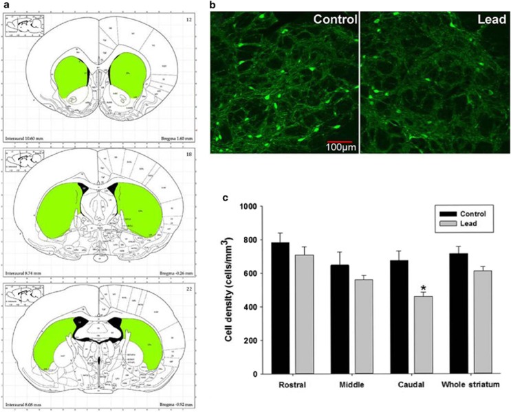
Figure 4.
Parvalbumin-positive cell density in the striatum of control and Pb2+ exposed rats. (a) Representative striatal regions used for PVGI cell counting (rostral, middle and caudal). (b) Immunofluorescence confocal imaging of PVGI in the caudal striatum from control and Pb2+ exposed animals. (c) PVGI cell density results for the striatum. While there was no overall effect of Pb2+ in the whole striatum when rostral, middle and caudal regions were averaged, we observed a significant decrease of PVGI in the caudal striatum of Pb2+ animals relative to controls when regions were analyzed individually. Data are represented as the mean (cells)±s.e.m. *, significantly different from control.

