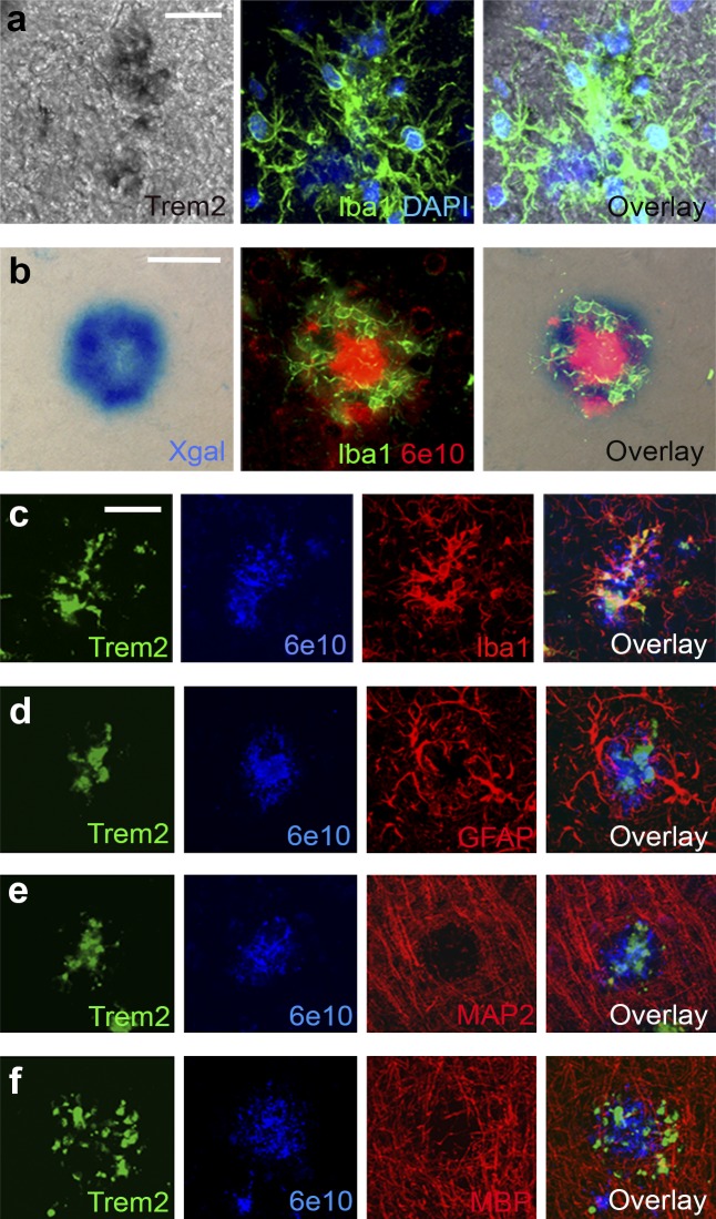
Figure 2.
TREM2 is expressed in plaque-associated myeloid cells. (a) In situ hybridization with TREM2 probes colocalized with Iba1 (n = 2). (b) X-gal staining of brain tissue from 4-mo-old APPPS1;Trem2LacZ/+ mice colocalized with fluorescent IHC for Iba1 and 6E10 (n = 3). (c–f) Confocal microscopy was used to assess TREM2 colocalization with 6E10+ plaque-associated myeloid cells (c; Iba1), astrocytes (d; GFAP), neurons (e; MAP2), or oligodendrocytes (f; MBP; n = 8). At least two independent experiments were performed for all analyses. Bars: (a) 20 µm; (b–f) 50 µm.

