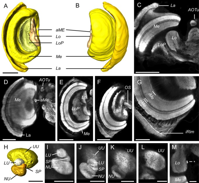
Figure 4.

Anatomy of the visual neuropils. A,B: Surface reconstructions of the optic lobe neuropils viewed from posterior (A) and anterior (B). They comprise the lamina (La), the medulla (Me), and accessory medulla (aMe), the lobula (Lo), and the lobula plate (LoP). C–G: Synapsin immunofluorescence in single confocal sections of the optic lobe. C: A horizontal section showing all four major optic lobe neuropils (La, Me, Lo, LoP) together with the anterior optic tubercle (AOTu) in the midbrain. D–F: Frontal sections at increasing depths from anterior to posterior, beginning at a plane tangential through the lamina (La in D) and reaching the optic stalk (OS in F). G: The inner rim (iRim) of the lamina is a thin layer on its inner surface that is defined by intense synapsin immunofluorescence; it is also visible in C,D,F. Synapsin immunostaining also reveals the laminated structure of the medulla with two main subdivisions, the outer and inner medulla (oMe, iMe). H: Surface reconstruction of the AOTu from an oblique anterior view, showing the four component neuropils: the upper unit (UU), lower unit (LU), strap (SP), and the nodular unit (NU). I–L: Synapsin immunofluorescence in the AOTu in frontal confocal sections at increasing depths from anterior (I) to posterior (L). M: A small neuropil (asterisk) positioned at the medial margin of the lobula which may be homologous to the D. plexippus optic glomerular complex. Scale bars = 200 μm in A–F; 100 μm in G; 50 μm in H–M.
