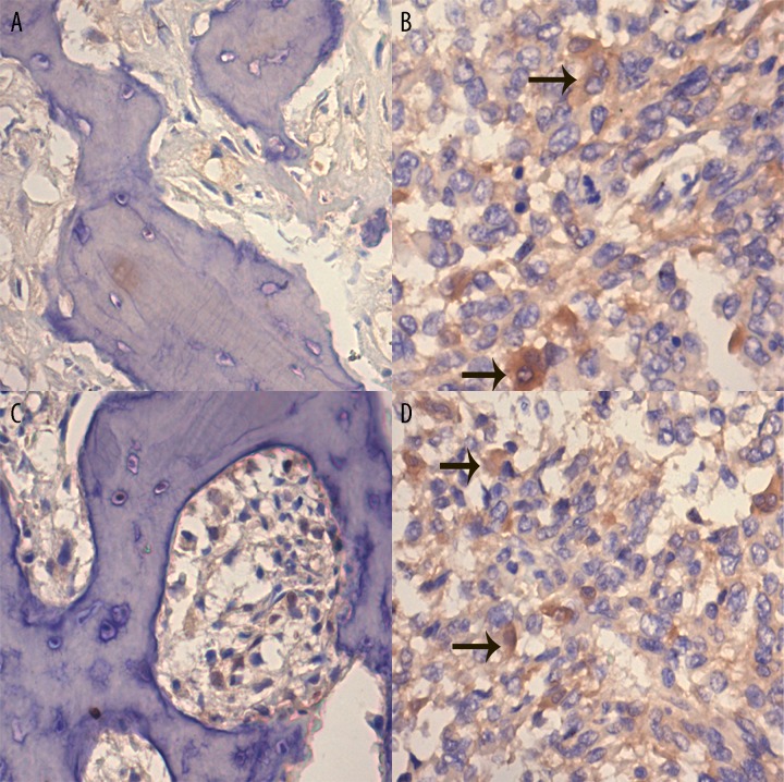
Figure 1.
Representative expression of Src and p-Src in osteosarcoma and osteochondroma samples. The sections were counterstained with hematoxylin and the positive staining is brown (indicated by black arrow). (A) immunohistochemistry of Src in osteochondroma specimen (×200). (B) immunohistochemistry of Src in osteosarcoma specimen (×200). (C) immunohistochemistry of p-Src in osteochondroma specimen (×200). (D) immunohistochemistry of p-Src in osteosarcoma specimen (×200).

