
Fig. 6.
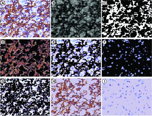
An experimental result from the present color-filtering method. (A) a representative IHC tissue image stained for CK; (B) the 8-bit BN image highlighting the contrast between DAB-stained tissues and hematoxylin background. The dark areas are the DAB-stained tissues, and the remaining areas are the Hematoxylin-stained tissues and background; (C) the brown-filter image with only two values: 0 or 1, multiplying by (A) to extract (D) a DAB-only-image containing brown DAB-stained tissues with black background and (E) a De-DAB-image; (F) the Hematoxylin -only-image containing blue Hematoxylin -stained tissues with black background; and (G) the Background image which is averaged to set as the back color of (D) and (F) to make (H) a DAB-stained-image and (I) a Hematoxylin -stained-image.