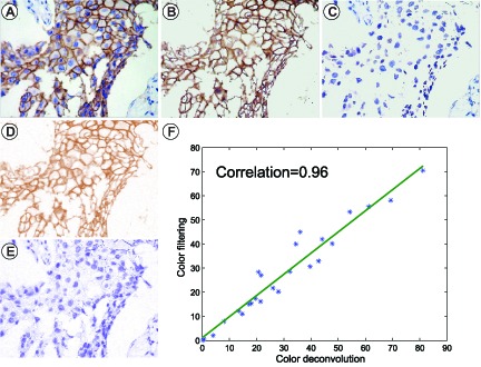
Fig. 8.

Positive color selection compared to color deconvolution analysis. (A) a representative immunohistochemical image stained for CK7; (B)(C) the DAB-stained-image and the Hematoxylin-stained-image obtained by color-filtering method showing the same values with original image, respectively; (D)(E) the DAB-stained-image and the Hematoxylin-stained-image obtained by color deconvolution method showing different RGB values with original image, respectively; and (F) Percentages of DAB-stained area display a strong association with results obtained from color deconvolution method.
