
Fig. 5.
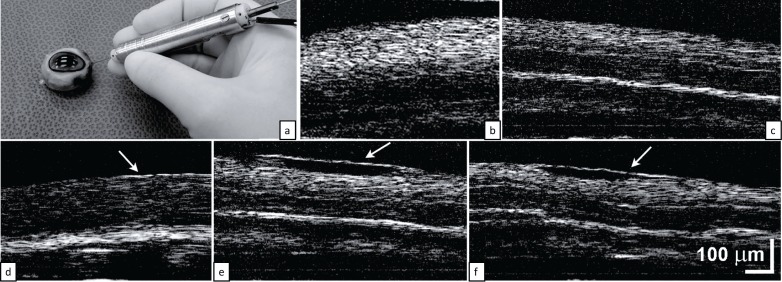
(a–c) The OCT probe with approximately 2mm scan length is capable of imaging retina through vitreous in an intact cadaver goat eye. (d–f) Ex vivo goat retina was used to enable application of artificial membranes. Nonuniform (d) tight adherence, (e) loose adherence, or (f) retinal contraction developed. Scale bars indicate the length of the images.