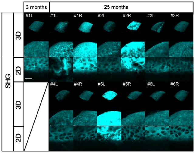
Fig. 8.
Results of 3D/2D image processing of the acquired SHG images of the medial condyle of the femur in 3-month-old mice (left knee; L) and 25-month-old mice (left and right knee; L and R, respectively). The upper two panels show volume rendering (3D) SHG images and their magnification. The lower panels show snapshot (2D) SHG images, scale bar: 25 μm.

