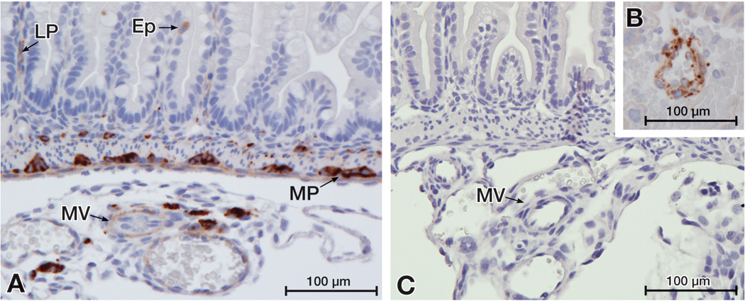
Figure 2. VEGF localizes to cells of the intestinal epithelium (Ep), the lamina propria (LP) and the myenteric plexus (MP) in the neonatal mouse small intestine.
The small intestine of day 1 dam-fed pups were immunostained for VEGF. (A) Positive staining shown in mesenteric vessels (MV) as well as in the LP, MP and few single cells of the Ep. (B) The specificity of VEGF staining was confirmed by positive staining of mouse placental vascular cells (B, positive control) and by the absence of staining in intestinal tissues when the primary antibody was omitted (C, negative control).

