
Figure 2. AngII-induced vasoconstriction was increased in SMC-HIF-1α-KO MA.
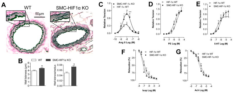
(A) Elastica van Gieson staining of mesesenteric arterery (MA) cross-sections, and (B) quantification of MA wall thickness and wall thickness/radius ratio. (C) Secondary MA function was examined with wire myograph. Cumulative concentration response curves to (C) AngII, (D) PE, (E) 5-HT, (F) Isop and (G) Ach (10−10–3×10⁻⁶ M for Ang-II and 10−8–3×10⁻⁵ M for the other agents). Data are presented as mean±S.E.M. ***p<0.001 vs. HIF-1α-WT; n=7 per group.