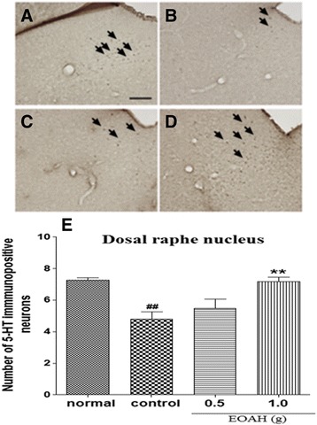
Figure 2.

Effect of inhalation of EOAH on 5-HT expression in the dorsal raphe nucleus in mice subjected to the FST. Photographs represent the distribution of 5-HT-immunoreactive neurons in the dorsal raphe nucleus of normal (A, n = 8), control (B, n = 9), EOAH 0.5 g (C, n = 9) and EOAH 1.0 g (D, n = 9) groups. The number of 5-HT immunostained neurons among the groups (E) was analyzed by one-way ANOVA post-hoc Newman-Keuls test. Each value represents the mean ± S.E.M. ## p < 0.01 compared to the normal group, **p < 0.01 compared to the control group. Sections were cut coronally at 30 μm and the scale bar represents 200 μm. Arrowheads indicate 5-HT immunopositive neurons.
