
Figure 4.
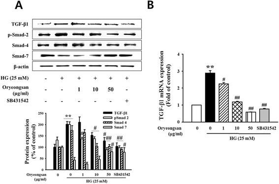
Effect of Oryeongsan on the relative levels of TGF-β 1 and Smads. (A) The protein bands detected by western blotting, β-actin was used as the internal standard in each sample. (B) real-time PCR showing mRNA levels of TGF-β in Oryeongsan-treated and HG-stimulated rat mesangial cells. Each value represents the means ± S.E. of five independent experiments. **p < 0.01 vs. control; #p <0.05, ##p < 0.01 vs. HG alone.