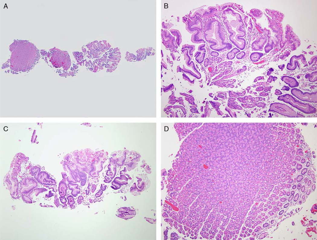
FIGURE 1.
A biopsy of a small gastric body polyp found during upper endoscopy of a JPS patient. The biopsy has both adjacent flat mucosa and fragments of the polyp (A). The polyp (B and C) has foveolar hyperplasia arising in oxyntic mucosa. There are reactive epithelial changes in the foveolar epithelium and scant plasma cells and lymphocytes in lamina propria. The flat mucosa (D) is histologically unremarkable. The polyp has the morphologic features of HP. As it is arising in gastric mucosa without significant pathology and in a JPS patient, this polyp is diagnosed as a gastric JP.

