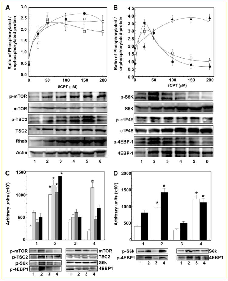
Fig. 4.
8-CPT concentration-dependent activation of mTOR signaling in 1-LN prostate cancer cells. Panel A: Changes in levels of p-mTOR (●), p-TSC2 (○) and Rheb protein (□) in 1-LN cells treated with varying concentrations of 8-CPT. Representative immunoblots of p-mTOR, p-TSC2 and Rheb along with immunoblots of the respective protein loading controls are shown below the diagram. 8-CPT-induced changes are shown as ratio of phosphorylated/unphosphorylated protein or actin in arbitrary units as the mean ± SE from four to five individual experiments. Panel B: Changes in phosphorylation of S6K (●), eIF4E (▲), and 4EBP-1 (□) in 1-LN cells stimulated with varying concentration of 8-CPT. Representative immunoblots of p-S6K, p-eIF4E and p-4EBP-1 along with the respective protein loading controls are shown below the diagram. 8-CPT-induced changes are shown as the ratio of phosphorylated/unphosphorylated protein, mean ± SE, from four to six individual experiments. Panel C: Inhibition of 8-CPT-induced increase in phosphorylation of mTOR (□) TSC2 (
 ), p-S6K (
 ), and 4EBP (■) by PI 3-kinase and mTOR inhibitors. The set of bars are: (1) buffer; (2) 8-CPT (50 μM); (3) LY294002 (20 μM/20 min) then 8-CPT (50 μM); and (4) rapamycin (100 nM/15 min) then 8-CPT (50 μM). Representative immunoblots of p-mTOR, p-TSC2, pS6K and 4EBP1 along with their protein loading controls are shown below the bar diagram. Changes in phosphorylation levels are shown in arbitrary units as the mean ± SE from four individual experiments. Panel D: Down regulation of 8-CPT-induced phosphorylation of S6K (□) and 4EBP1 (■) in 1-LN cells transfected with Epac1 dsRNA. The bars are: (1) lipofectamine + buffer; (2) lipofectamine + 8 + CPT; (3) lipofectamine + Epac1 dsRNA (100 nM) then 8-CPT; and (4) scrambled dsRNA (100 nM) + 8-CPT. Representative immunoblot of p-S6K and p-4EBP1 are shown below the bar diagram. Changes in phosphorylation levels are shown in arbitrary units as the mean ± SE from three individual experiments. *Values significantly different at the 5% levels comparing the buffer controls, inhibitor-treated or Epac1 dsRNA transfected cells to 8-CPT-treated cells.

