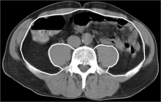
Fig 2. The image illustrates how the area of visceral adipose tissue (VAT) within the abdominal cavity (white line) was determined by CT scanning.

Pixels with the density of adipose tissue between −30 and −190 Hounsfield Units (HU) were included in the VAT area calculated automatically by the CT software. In the illustrative case, the VAT area is 110 cm2.
