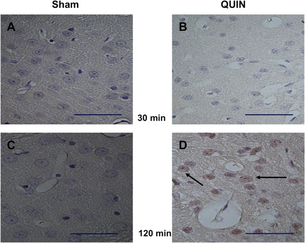
Fig 4. Histochemical labeling of RAGE-positive cells.
Peroxidase-based immunohistochemical staining of RAGE-positive cells in striatal coronal sections (40X) of Sham (A and C)- and QUIN (B and D)- treated animals at 30 and 120 min post-lesion, respectively (Bar size 100 μm). In D, a prominent reactivity of cells to RAGE (indicated by arrows) is observed.

