
Figure 3.
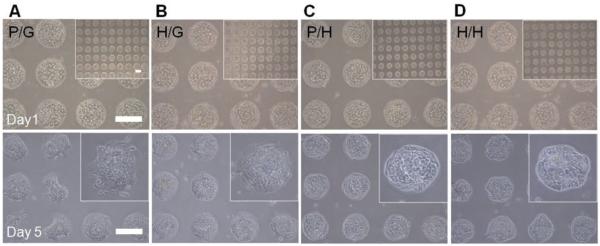
Culturing primary rat hepatocytes on the four kinds of gel microstructures of varying components. A) Bright-field microscopic images of hepatocytes cultured on PEG microwells on the glass surface (P/G). B) Heparin gel microwells on the glass surface (H/G). C) PEG microwells on soft heparin gel layer (P/H). D) Heparin microwells on soft heparin gel layer (H/H) at day 1 and day 5. Scale bar = 200 μm.