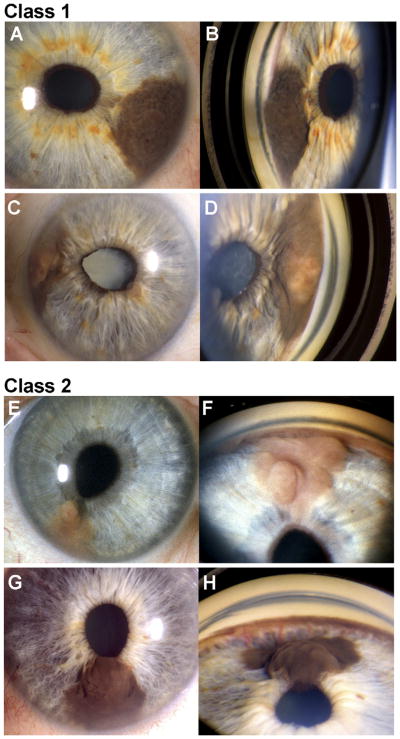
Figure 1.
Examples of class 1 and class 2 iris melanomas. Paired slit-lamp photographs (left panels) and goniophotographs (right panels) of 2 class 1 iris melanomas (A–D) and 2 class 2 iris melanomas (E–H). Patient NB018 (A–B) presented with a dark brown tumor arising from the iris stroma and extending into the angle and causing slight peaking of the pupil (not a diagnostic criterion for iris melanoma). Patient NB006 (C, D) presented with a variably pigmented iris melanoma with angle involvement, peaking of the pupil, and secondary cataract. Patient NB206 (E, F) presented with a minimally pigmented, multilobular iris melanoma with angle involvement and peaking of the pupil. This tumor recurred with vitreous seeding 71 months after I-125 plaque radiotherapy. Patient NB138 (G, H) presented with a dark brown iris stromal tumor with no angle involvement. Prominent feeder vessels emanating from the angle could be seen on gonioscopy (H). Despite the lack of angle involvement, the tumor grew substantially on observation, so I-125 plaque radiotherapy was performed.

