Figure 3.
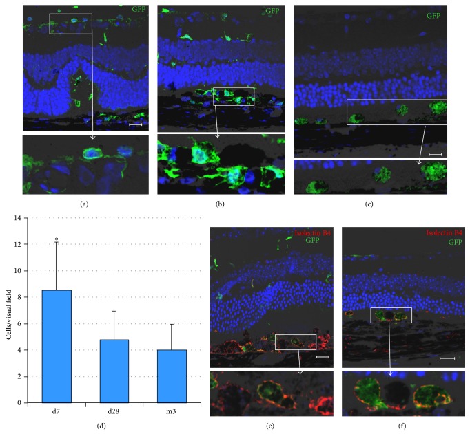
Longitudinal follow-up of the fate of Lin−BMCs at different time points post intravitreal transplantation. Immunofluorescence analysis of retinas after intravitreal GFP+Lin−BMCs delivery showed transplanted cells (green) on the 7th day (a, b) and at 3 months after injection (c). Quantitative analysis of the total number of incorporated Lin−BMCs per eye section (n = 5/time point) (d). Coreactivity of GFP (green) and Isolectin-B4 (red) by fluorescent lectin staining showed that transplanted GFP+Lin−BMCs locally differentiated into Isolectin-B4-positive macrophages (inserts) in Lin−BMC-injected eyes on the 7th day after transplantation (e) and at 3 months after injection (f). Scale bar = 20 μm. * P < 0.05 for day 7 versus other time points.

