Figure 5.

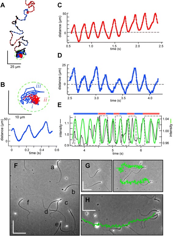
Release from transient attachment allows change in orientation and trajectory. (A) Tracking a transiently attached sperm. Duration 6 s, sampling 100 frames per second (fps), medium HSB. Right cheek (RCh, red) and left cheek (LCh, blue) orientations are indicated. Green circle encloses initial 4 s period of attachment. (B-D) Time courses of changes in flagellar location (from linescans perpendicular to the flagellar axis at approximately 15 μm from the flagellar origin) during attachment. (B) Inset shows expanded view of track during attachment, divided into segments of LCh (i, iii) and RCh (ii) orientation. Time course for segment i. (C, D) Time courses for subsequent RCh (ii) and LCh (iii) attachment. For C and D, images were first ‘despun’ by rotation of 0.1 degrees per frame. (E) Time courses of centroid region-of-interest intensities (black) and best-fit sine functions (green) during subsequent free-swimming of the released sperm. Bars indicate periods of LCh (blue) and RCh (red) orientation. (F) Coexistence of various swimming behaviors in a single 240 × 350 μm field. Medium HSB. Lower case letters indicate cells showing: (a, b) linear swimming by two unattached sperm - sperm a1 exits at t = 0.2 s, sperm a2 enters at t = 3.5 s; (c) tethered spinning of an attached sperm duo; (d) linear swim by an unattached, headless sperm; (e) transient linear swim terminated by attachment at t = 3.5 s; (f) transient tether followed by linear free swim (the cell examined in A-E). (G, H) Tracking of cells from other fields of this same preparation, examined under the same conditions as in panel A except for use of medium HS rather than HSB. (G) Two cells at the start (left) and end (right) of a 2 s linear swim. (H) A trio of attached cells at the end of a 2 s linear swim. Swimming tracks are green. Scale bars, 50 μm.
