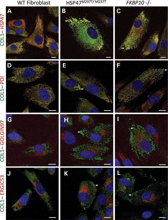
Figure 8.
Type I procollagen is present in ER-related abnormal vesicles in OI-mutant cells. Immunofluorescence of COL1 (green) and HSP47 and cell compartments (red): HSP47 (A–C), PDI for ER (D–F) Golgin97 for Golgi apparatus (G–I) and ERGIC53 for ER–Golgi intermediate compartment (J–L). Control (A, D, G and J), HSP47M237T/M237T (B, E, H and K) and FKBP10−/− cells (C, F, I and L). White arrows identify vesicles accumulating HSP47 and type I procollagen. Bars represent 10 µm.

