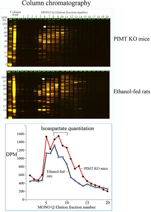
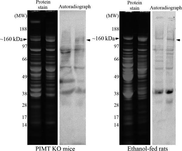
Figure 3. Enrichment and partial purification of isoaspartate damaged proteins.
(A) Liver cytosolic proteins from PIMT KO mice or ethanol-fed rats were enriched through binding and elution from a MONO Q column. Proteins were stained with colloidal Coomassie (upper panel). The start of elution of the ~160 kDa protein doublet in fraction 7 is marked with an arrowhead. The level of isoaspartate damage in each fraction was quantified (lower panel). (B) Peak column fractions 7-10 were concentrated, fractioned, and then resolved by 1D PAGE. Proteins were stained with colloidal Coomassie, or radiolabelled by PIMT using 3H-SAM, and radiolabelled proteins visualised by autoradiography. Co-distribution of protein staining and isoaspartate radiolabelling was evident for the ~160 kDa protein for both PIMT KO mice and ethanol-fed rats (marked with arrowheads).


