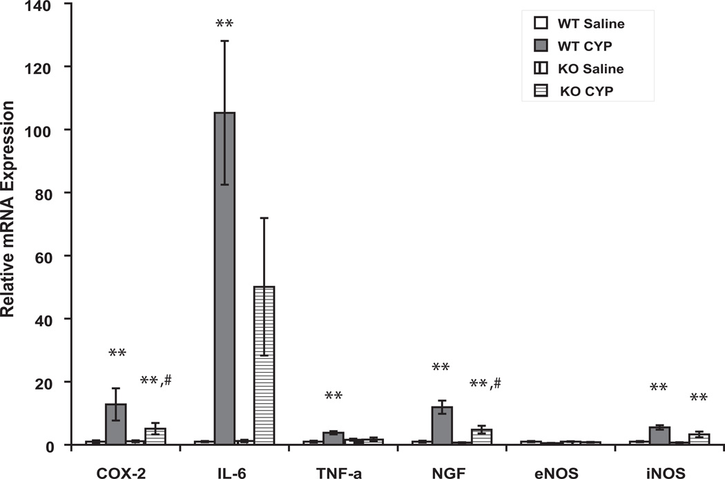
Figure 4.
Expression of mRNAs of pro-inflammatory compounds 3 hours after CYP. Expression of each gene is normalized to abundance of mRNA for L19, a constitutively-expressed ribosomal protein (house-keeping gene) in the same sample. Fold changes of expression of each gene in CYP-treated WT and saline- and CYP-treated FAAH KO mice were compared to the corresponding gene expression in saline-treated WT animals (set as 1). n = 6–8 for each treatment. *, ** indicate p <0.05 and 0.01, respectively, CYP vs saline for each genotype. # indicates p < 0.05 vs CYP-treated WT mice.

