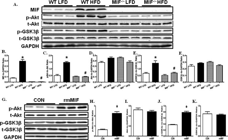
Fig. 4.
MIF deficiency ameliorated HFD-induced cardiomyocyte Akt pathway activation. A: Representative gel blots depicting levels of MIF, pAkt, Akt, pGSK-3β, GSK-3β, and GAPDH (as loading control) using specific antibodies; B: MIF expression; C: Akt phosphorylation (Ser473, p-Akt-to-Akt ratio); D: total Akt expression; E: GSK-3β phosphorylation (Ser9, p- GSK-3β -to- GSK-3β ratio); F: total GSK-3β expression. Mean ± SEM, *p < 0.05 vs. WT LFD group, #p < 0.05 vs. WT HFD group. G: Representative gel blots showing that recombinant mouse MIF (rmMIF, 50 ng/ ml, 6 hr) induces Akt pathway activation in cardiomyocytes isolated from WT mice; H: Akt phosphorylation (Ser473, p-Akt-to-Akt ratio); I: total Akt expression; J: GSK-3β phosphorylation (Ser9, p- GSK-3β -to- GSK-3β ratio); and K: total GSK-3β expression. Mean ± SEM, *p < 0.05 vs. control (CON) group.

