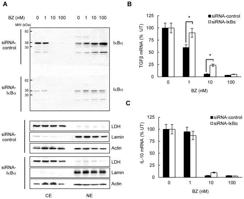
Figure 2. TGFβ1 inhibition is mediated by IκBα, while IL-10 inhibition is IκBα-independent.
(A) Western blotting of cytoplasmic (CE) and nuclear (NE) extracts prepared from Hut-78 cells transfected with control non-silencing (top gel) and IκBα specific (bottom gel) siRNA, incubated 24 hours with increasing BZ concentrations, and analyzed by using IκBα antibody. Both gels were transferred to a membrane and exposed together at the same time. The purity of cytoplasmic and nuclear fractions was monitored by using lactate dehydrogenase (LDH) and lamin B antibodies. To confirm equal protein loading, the membranes were stripped and re-probed with actin antibody. (B) TGFβ1 and (C) IL-10 mRNA levels in Hut-78 cells transfected with control and IκBα siRNA, and treated 24 hours with increasing BZ concentrations. The values represent the mean +/−SE of four experiments; the asterisks denote a statistically significant (p<0.05) change compared to cells transfected with control siRNA.

