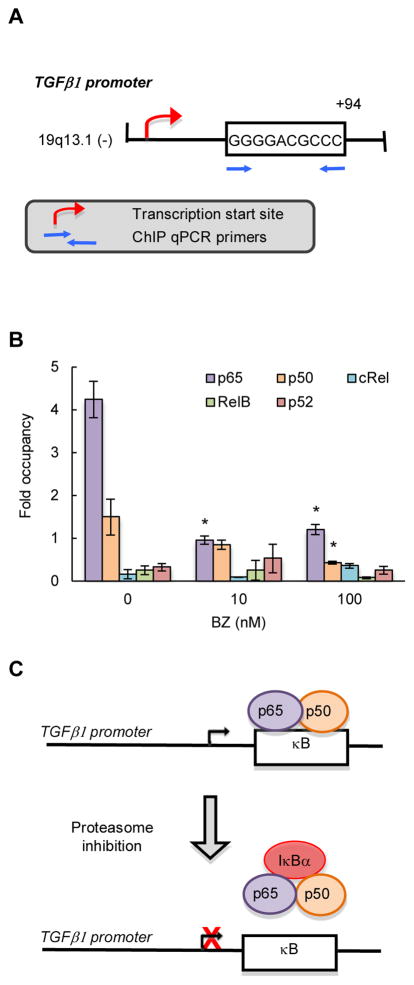
Figure 3. TGFβ1 is regulated by NFκB canonical pathway in CTCL cells.
(A) Schematic illustration of NFκB binding site in human TGFβ1 promoter, and the ChIP primers used in the ChIP assay. (B) Recruitment of NFκB p65, p50, cRel, RelB and p52 subunits to TGFβ1 promoter in Hut-78 cells treated 24 hours with 0, 10 and 100 nM BZ was analyzed by ChIP and quantified by real time PCR. The data are presented as the difference in occupancy of each protein between the TGFβ1 promoter and the IGX1A (SA Biosciences) negative control locus, and represent the mean +/−SE of four experiments. Asterisks denote a statistically significant (p<0.05) change compared to control (no BZ) cells. (C) Model of the regulation of TGFβ1 transcription in CTCL cells.

