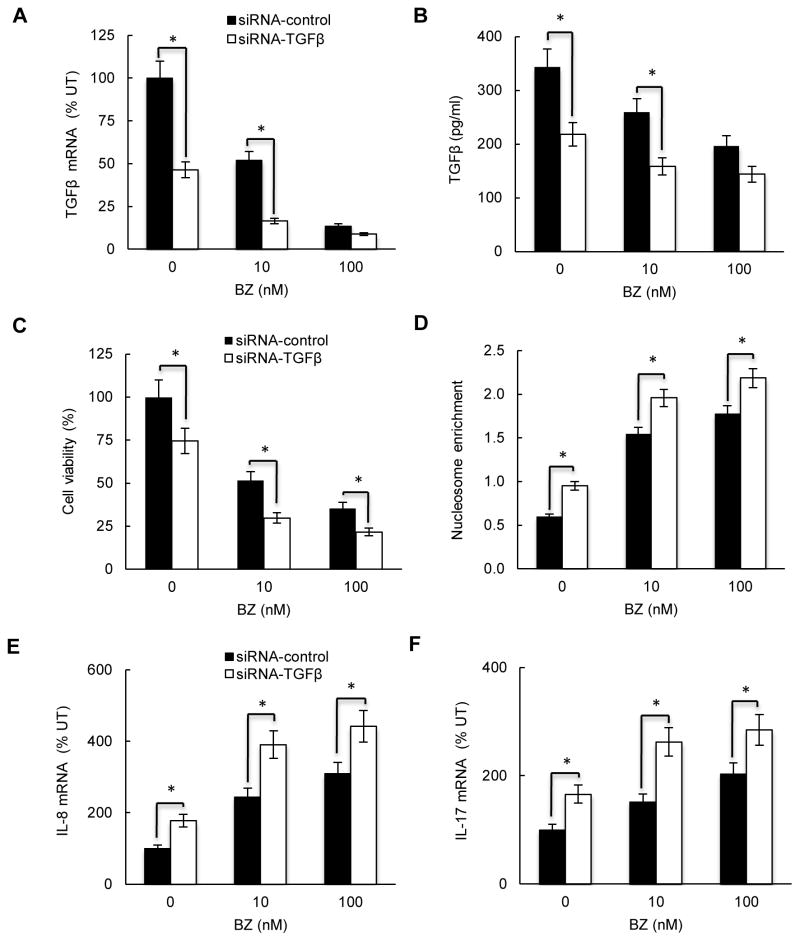
Figure 5. TGFβ1 suppression decreases viability, and increases pro-inflammatory gene expression in CTCL cells.
HH cells were transfected with control (full columns) or TGFβ1 specific siRNA (empty columns), treated 24 hours with 0, 10 and 100 nM BZ, and analyzed for TGFβ1 mRNA expression by real time RT-PCR (A), and for TGFβ1 release by ELISA (B). (C) Cell viability measured by Trypan Blue exclusion, and (D) apoptosis analyzed by the cytoplasmic nucleosome enrichment assay in HH cells transfected with control or TGFβ1 specific siRNA. Real time RT-PCR analysis of IL-8 (E) and IL-17 (F) mRNA levels in HH cells transfected with control (full columns) or TGFβ1 specific siRNA (empty columns) and treated 24 hours with 0, 10 and 100 nM BZ. The values in Figs. 5A-F represent the mean +/−SE of four experiments. Asterisks denote a statistically significant (p<0.05) change compared to cells transfected with the corresponding control siRNA.

